Microscopes magnify small objects that cannot be seen by the naked eye to observe them at a cellular level. Microscopes are used in different fields for different purposes, right from studying cells for educational purposes to identifying bacteria and viruses to diagnose and treat diseases to even identifying evidence and solving forensic cases. The modern microscope has come a long way since the Dutch optician Zacharias Janssen built the first simple microscope in the late 1500s.
There are many types of microscopes ranging from compound microscopes to simple microscopes to electron microscopes.
Origin of an inverted microscope
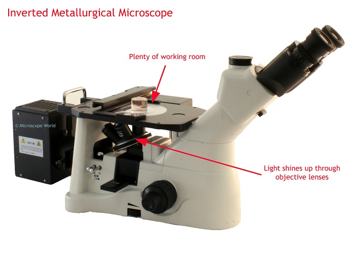
One of the popular techniques to view live cell imaging is through an inverted microscope. Invented by John Smith in 1850, he flipped the traditional design to build an inverted microscope. The objective lenses of an Inverted microscope are located below the Stage so that light travels downward from the specimen to the objective lenses below. The advantage of this device is that large samples can be magnified without cutting them into thin slices.
Though inverted microscopes were primarily used in the metal industry to examine metal samples for composition, coatings, grain size, and defects, later in the 20th century, scientists and microbiologists started to use the apparatus more.
Parts of an inverted microscope
- The specimen is kept over the Stage
- The specimen is held in position by Stage Clips
- An Arm that holds the optical and mechanical parts of the microscope.
- An Objective Lens to magnify the images. It moves along the vertical axis.
- Dual concentric knobs to focus and fine-tune the microscope.
- A Nosepiece to hold the objective lens.
- A Condenser lens concentrates the light on the specimen.
- A Digital Camera to record or capture the image of the specimen.
The function of an inverted microscope
As mentioned above, the transmitted light source and condenser are found on the top of the Stage, pointing downwards toward the Stage. Observations are made through the bottom of the cell culture vessel with the objectives located below the Stage pointing upwards.
Common applications of an inverted microscope
- Probably, the most important application of an inverted microscope is to observe the living cells and tissues in their natural state. As part of certain diagnostic tests, the inverted microscope is also used to observe drug sensitivity (MODS) in certain samples.
- As a diagnostic tool, it can be used to detect Phytophthora species in fungal cultures, for example.
- In parasitology, diagnosing nematodes by observing vermiform nematodes in extraction specimens is useful.
- Microscopes are used in diagnosis methods to examine Mycobacterium tuberculosis parasites.
- Inverted microscopes are used in micromanipulation processes.
Benefits of using an inverted microscope
Some of the significant advantages of using an inverted microscope are mentioned below.
- You can use specimens of varying sizes and weights, making the inverted microscope versatile and ideal for industrial use.
- Unlike traditional microscopes, sample preparation is not laborious and requires no expertise. You can place the sample over the Stage and magnify it.
- A live sample can be observed over time which allows for studying the cells at different stages of division.
It is especially important to consider the advantages that you might get over a conventional microscope when working with live specimens or tissues that require additional space or a larger working distance. For details visit our website at RS online.


